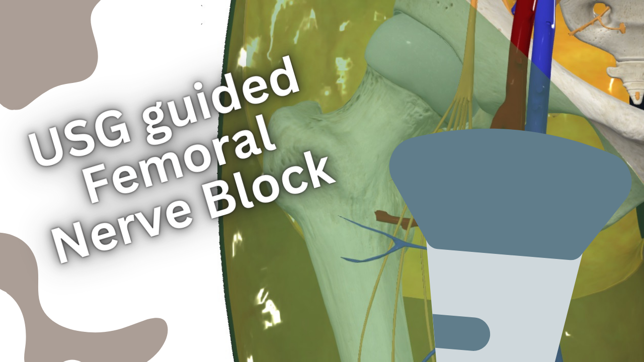
USG-Guided Femoral Nerve Block

Ultrasound-Guided Femoral Nerve Block: Precision in Lower Limb Anesthesia & Pain
Introduction
USG-guided femoral nerve block (FNB) is a highly effective regional anesthesia technique for surgeries and pain relief involving the anterior thigh, knee, and hip. This method, which uses real-time ultrasound imaging to accurately target the femoral nerve, offers numerous benefits including improved patient safety, enhanced analgesia, and reduced reliance on systemic opioids. This article provides a detailed exploration of the femoral nerve block, its anatomical basis, procedural technique, clinical applications, and advantages.
Anatomy of the Femoral Nerve
The femoral nerve is one of the largest nerves in the body and is essential for the motor and sensory innervation of the anterior thigh. Understanding its anatomy is crucial for performing effective regional anesthesia, particularly in procedures such as femoral nerve blocks. This article provides a comprehensive overview of the femoral nerve’s anatomy, its branches, and its clinical significance in regional anesthesia.
Origin and Course of the Femoral Nerve
Origin
- Roots: The femoral nerve arises from the posterior divisions of the L2, L3, and L4 spinal nerves.
- Plexus: It is a major branch of the lumbar plexus, which is formed within the psoas major muscle.
Course
- Psoas Major Muscle: The femoral nerve descends through the psoas major muscle and emerges from its lateral border.
- Pelvis: It travels down between the psoas major and iliacus muscles.
- Inguinal Ligament: The nerve passes under the inguinal ligament, entering the thigh.
- Femoral Triangle: It enters the thigh within the femoral triangle, a region bounded by the inguinal ligament (superiorly), the sartorius muscle (laterally), and the adductor longus muscle (medially).
Divisions
Upon entering the thigh, the femoral nerve divides into several branches that provide motor and sensory innervation to the anterior thigh and lower leg.
Branches of the Femoral Nerve
Muscular Branches
- Iliacus Muscle: Before passing under the inguinal ligament, the femoral nerve supplies the iliacus muscle.
- Pectineus Muscle: It innervates the pectineus muscle, which assists in hip flexion and adduction.
- Sartorius Muscle: The nerve innervates the sartorius muscle, involved in hip and knee flexion.
- Quadriceps Femoris: The femoral nerve provides branches to the quadriceps femoris group, which includes the rectus femoris, vastus lateralis, vastus medialis, and vastus intermedius muscles. These muscles are crucial for knee extension.
Sensory Branches
- Anterior Cutaneous Branches: These branches arise in the femoral triangle and provide sensory innervation to the skin of the anterior and medial thigh.
- Saphenous Nerve: This is the longest branch of the femoral nerve. It descends along the medial side of the leg to provide sensory innervation to the skin over the medial side of the knee, leg, and foot.
The Technique of USG-Guided Femoral Nerve Block
Patient Preparation
- Positioning: The patient is positioned supine with the leg slightly abducted and externally rotated.
- Equipment: A high-frequency linear ultrasound probe is used to visualize the femoral nerve and surrounding structures.

Procedure
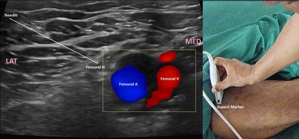
- Probe Placement: Place the ultrasound probe transversely in the inguinal crease to obtain a short-axis view of the femoral nerve.
- Identification of Structures: Identify the femoral artery and vein, with the femoral nerve located lateral to the artery. The nerve typically appears as a hyperechoic (bright) structure.
- Needle Insertion: Using an in-plane technique, insert the needle from lateral to medial. Continuous ultrasound visualization ensures precise needle guidance.
- Confirmation of Needle Placement: The needle tip should be adjacent to the femoral nerve, which can be confirmed by slight movement of the nerve or tissue around it upon needle contact.
- Anesthetic Injection: Slowly inject the local anesthetic while observing its spread around the nerve. Adequate spread typically encases the nerve, confirming successful blockade.
Clinical Applications of USG guided femoral nerve block
Surgical Anesthesia
- Knee Surgery: Commonly used for procedures such as total knee arthroplasty (TKA) and anterior cruciate ligament (ACL) reconstruction.
- Hip Surgery: Provides effective analgesia for hip fracture repair and other hip surgeries.
- Thigh Surgery: Useful for operations involving the quadriceps muscle and other anterior thigh structures.
Postoperative Analgesia
FNB offers prolonged pain relief following lower limb surgeries, significantly reducing the need for systemic analgesics and enhancing patient comfort during the postoperative period.
Emergency and Trauma Settings
In emergency settings, FNB can provide rapid and effective pain relief for femoral fractures and other acute lower limb injuries.
Advantages of Ultrasound Guidance
Enhanced Accuracy
Ultrasound guidance allows for precise identification of the femoral nerve and accurate needle placement. This reduces the likelihood of complications associated with blind or landmark-based techniques, such as vascular puncture or intraneural injection.
Improved Patient Safety
By visualizing the needle and nerve in real-time, the risk of nerve injury and other complications is minimized. Additionally, ultrasound guidance reduces the volume of local anesthetic required to achieve effective blockade, further enhancing safety.
Superior Analgesia
Ultrasound-guided FNB provides more consistent and reliable analgesia compared to traditional techniques. The precise delivery of local anesthetic ensures optimal nerve blockade, resulting in superior pain control.
Reduced Opioid Consumption
Effective regional anesthesia reduces the need for systemic opioids, lowering the risk of opioid-related side effects, dependency, and overdose. This is particularly beneficial in the context of the current opioid epidemic.
Potential Challenges of USG-guided Femoral Nerve Block
Learning Curve
Proficiency in ultrasound-guided techniques requires dedicated training and practice. Anesthesiologists must develop skills in ultrasound imaging, needle guidance, and interpretation of sonographic anatomy. Simulation training and hands-on workshops can help mitigate this learning curve.
Equipment Availability
High-quality ultrasound machines and appropriate probes are essential for successful ultrasound-guided FNB. Access to such equipment may vary between institutions, potentially limiting the widespread adoption of these techniques.
Anatomical Variations
Variations in femoral nerve anatomy can pose challenges to successful blockade. Anesthesiologists must be adept at identifying and adapting to these variations to ensure effective anesthesia.
Conclusion
Ultrasound-guided femoral nerve block is a transformative technique in regional anesthesia, offering unparalleled precision, safety, and efficacy for lower limb surgeries and pain management. Its ability to provide superior pain relief, reduce opioid consumption, and improve patient outcomes underscores its importance in modern anesthetic practice. Despite challenges such as the learning curve and equipment requirements, the overall benefits of ultrasound-guided FNB make it a valuable addition to the anesthesiologist’s toolkit. As technology advances and training becomes more standardized, the role of ultrasound-guided nerve blocks will continue to grow, further enhancing the quality of patient care.

